

 CALL THE LOCATION NEAREST YOU
Wisdom Teeth X-Rays: How Oral Surgeons Assess Your Case
Why Wisdom Teeth X-Rays Matter Before Treatment
Wisdom teeth can sit partly or entirely beneath the gums, so a visual examination only tells part of the story. X-rays give an oral surgeon a view of the roots, surrounding bone, and nearby structures before treatment is considered. The images do not make the decision by themselves, but they provide the map needed for a safer, more individualized discussion.
An oral exam can reveal swelling, gum irritation, tenderness, drainage, and whether a wisdom tooth has erupted into the mouth. What it cannot reliably show is the full position of a tooth under bone, the shape of its roots, or its relationship to structures that are not visible from the surface. A dental image can show whether a wisdom tooth is tilted, trapped, or directed toward the second molar, and may reveal decay, changes in the surrounding bone, or a space that needs closer evaluation.
When Imaging Is Recommended
Imaging is commonly considered when wisdom teeth are developing, when a tooth is partly erupted, or when an examination raises questions about its position. A new scan may also be useful before planned removal, especially if earlier images are outdated or do not show the area clearly. The timing depends on age, symptoms, dental history, and what the clinician sees during the examination.
Pain is not a dependable guide to the condition of every wisdom tooth. Some people have significant inflammation with obvious discomfort, while others have an impacted tooth that remains quiet for a long time. That is why the decision to monitor or treat usually combines symptoms, examination findings, and imaging rather than relying on how the mouth feels on one particular day.
The Types of X-Rays Used to Evaluate Wisdom Teeth
Different images answer different clinical questions. A panoramic X-ray captures a broad view of the upper and lower jaws, including the wisdom teeth, neighboring molars, and portions of the surrounding bone, and is often a useful starting point when the question involves more than one wisdom tooth. Intraoral images place the detector inside the mouth and focus on a smaller region, helping clarify the crown of a tooth, the adjacent molar, or the bone around a specific area. Cone beam computed tomography, often called CBCT, produces three-dimensional views of the jaws and teeth and may be considered when the position of a tooth or its roots needs to be understood from more than one direction.
More than one type of image may be useful when a broad overview identifies a concern that needs closer inspection. A second scan should add information rather than simply repeat an image without a clinical purpose.
| Imaging Type | Main View | Typical Planning Value |
|---|---|---|
| Panoramic X-ray | Broad view of both jaws | Reviews overall tooth position and development |
| Intraoral X-ray | Focused view of a small area | Clarifies a tooth, root, or nearby molar |
| CBCT scan | Three-dimensional view | Examines spatial relationships from multiple angles |
The most suitable image is the one that answers the question at hand with an appropriate amount of detail. Your provider can explain why a particular scan is sufficient or why additional imaging would change the evaluation.
What Oral Surgeons Look for in the Images
An oral surgeon reads wisdom teeth images as a collection of relationships, not as an isolated picture of one tooth. The tooth's direction, root shape, eruption status, and surrounding structures all contribute to the assessment, with the goal of identifying present problems and anticipating technical issues that could affect monitoring or removal.
A wisdom tooth may be upright, tilted toward the second molar, angled toward the cheek or back of the jaw, or lying nearly horizontal, and its position can affect whether it has a realistic path into the mouth. An impacted wisdom tooth is blocked from fully emerging, while a partially erupted tooth has broken through only part of the gum; partially erupted teeth can be difficult to clean around.
Lower wisdom tooth roots may lie close to the canal that carries an important nerve, while upper wisdom teeth can be near the maxillary sinus, and the image also shows whether a wisdom tooth is pressing against or damaging the second molar. Images may also show changes around the crown, loss of tooth structure, decay in a neighboring molar, or alterations in the surrounding bone.
How X-Ray Findings Affect Your Treatment Plan
An X-ray is one part of a treatment conversation rather than an automatic instruction to remove every wisdom tooth. The plan may involve observation, improved cleaning, treatment of an active problem, or extraction, shaped by the tooth's position, the condition of nearby structures, the person's symptoms, and the risks of both action and inaction.
A wisdom tooth that is healthy, stable, and not causing a problem may be monitored rather than removed immediately, generally meaning periodic examinations and updated imaging when clinically appropriate. An impacted tooth may require an incision, removal of some surrounding bone, or sectioning of the tooth so it can be taken out in manageable pieces; the exact approach depends on depth, angle, root shape, and access. When a lower wisdom tooth appears close to a nerve canal, the surgeon may discuss the limits of the image and the possible risks in more detail, and additional three-dimensional imaging may be considered if it could clarify the relationship.
A referral to our oral surgery team may be appropriate when a tooth is deeply impacted, close to important anatomy, associated with a cyst or significant infection, or technically difficult to access.
What to Expect During Wisdom Teeth X-Rays
Dental imaging is usually a brief, noninvasive part of an evaluation. The staff will position you for the selected scan and may ask you to remove items that could interfere with the image. If you know you are pregnant, have difficulty standing still, or have concerns about the procedure, mention them before imaging begins.
Most wisdom teeth X-rays do not require fasting or special preparation. Follow any instructions from the office, arrive with relevant medical information, and remove jewelry or hair accessories around the head and neck if requested. It can also help to bring earlier images if they were taken at another office, since comparison may prevent unnecessary repeat imaging.
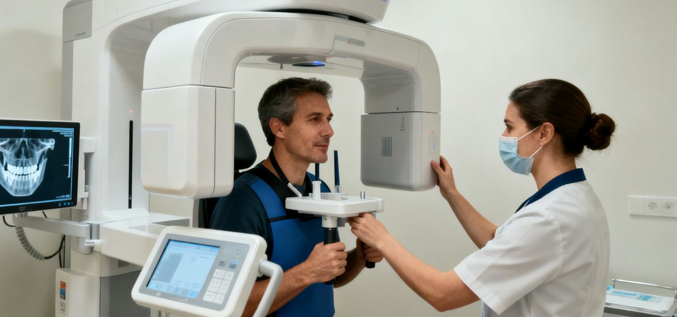
For a panoramic image, you will usually stand or sit at the machine and rest your chin on a support while the machine rotates around your head. During a CBCT scan, you may stand or sit while the scanner rotates around your head, capturing multiple views that are combined into a three-dimensional dataset. The positioning and exposure for a dental image are typically brief, although the complete appointment may take longer if several images are needed or if a consultation follows.
Understanding Radiation Exposure and Safety
Dental X-rays use ionizing radiation, so imaging is ordered when the expected clinical value justifies it. Modern dental offices aim to obtain the needed information with the lowest reasonable exposure and avoid repeating images without a clear reason. Providers select the smallest field and appropriate settings that can answer the clinical question, and digital systems allow images to be captured and reviewed efficiently while careful positioning helps avoid retakes.
Tell your dentist, radiographer, or oral surgeon if you are pregnant or might be pregnant before the scan, so the care team can consider timing, alternatives, and whether the image is needed now. The decision to image depends on whether the result could change diagnosis, treatment, or surgical planning; if a hidden tooth position or nearby structure could materially affect care, the information gained may outweigh the small radiation exposure involved.
Questions to Ask After Reviewing Your X-Rays
Seeing an X-ray can be confusing because the image contains more anatomy than most patients are used to interpreting. Ask the clinician to point out each wisdom tooth and explain what is relevant rather than trying to diagnose yourself from the screen. A good review should connect the image to your symptoms, examination, and choices.
Ask whether each tooth is upright, tilted, impacted, partially erupted, or fully erupted, and whether there is enough space for it. If removal is being discussed, ask what specific finding supports it and what the alternative would be; if monitoring is reasonable, ask how often the tooth should be checked and which symptoms should prompt a call. A clinician can also explain whether the image identified risks such as damage to a second molar, recurrent gum infection, decay, or a difficult relationship to nearby anatomy, and whether another type of imaging would add useful information to the plan. Our Frequently Asked Questions page covers many of these common concerns, and you can reach our team through the Contact page with additional questions.
Conclusion
Wisdom teeth X-rays help oral surgeons see what an examination cannot: the tooth's position, roots, surrounding bone, and relationship to nearby structures. Panoramic, intraoral, and three-dimensional images each have a different role, and the right choice depends on the clinical question. According to the American Dental Association, wisdom teeth can lead to problems if there isn't enough space for them to come through properly, which is why your dentist examines your mouth and takes an X-ray before recommending a course of treatment. Reviewing the findings with your provider can make the decision to monitor, investigate further, or remove a tooth more understandable and deliberate.
Frequently Asked Questions
Are wisdom teeth X-rays always necessary before removal?
Imaging is commonly used before removal because it helps show tooth position, roots, bone, and nearby anatomy. The type and extent of imaging depend on the examination and the questions the clinician needs to answer.
Can an impacted wisdom tooth cause no symptoms?
Yes. An impacted tooth may remain quiet, even though its position could affect nearby teeth or become difficult to manage later. Regular evaluation helps identify changes that symptoms alone might miss.
Which X-ray is usually used first for wisdom teeth?
A panoramic X-ray is often used for an overall view of the jaws and wisdom teeth. Focused intraoral images or CBCT may be considered when more detail is needed.
Does every impacted wisdom tooth need to be removed?
No. The decision depends on symptoms, tooth condition, nearby structures, cleaning access, and the risks of monitoring versus removal. Some teeth can be observed with appropriate follow-up.
How long does a wisdom teeth X-ray take?
The exposure itself is usually brief. The full visit may take longer if positioning, multiple images, a three-dimensional scan, or a consultation is needed.
Should I tell my dentist if I may be pregnant?
Yes. Tell the dental team before imaging so they can consider the timing and necessity of the scan. They can discuss the situation and any reasonable alternatives with you.
Can an X-ray show whether extraction will be difficult?
An image can reveal factors such as impaction, root shape, depth, and proximity to nerves or sinuses. These findings help the surgeon estimate complexity and plan the procedure, although no image predicts every detail with certainty.
About the Author
Oral Surgeons, PC has served the Des Moines, Iowa area for decades from offices in Des Moines, Ankeny, and Clive, with a surgical team that includes Dr. Johnson, Dr. Janulewicz, Dr. Marsh, Dr. Freml, Dr. Morio, Dr. Radich, Dr. Ridenour, Dr. Peters, and Dr. Schwarzkopf. The practice is a proud member of the American Association of Oral and Maxillofacial Surgeons and the American Dental Association, providing wisdom teeth evaluation, imaging, and extraction to patients throughout the Des Moines metro area, working closely with referring general dentists on complex or impacted cases.
Disclaimer
The information provided in this article is for educational purposes only and does not constitute professional medical or dental advice, diagnosis, or treatment recommendations. Whether imaging or extraction is appropriate for your wisdom teeth can only be determined through an in-person evaluation. Always consult your dentist or oral surgeon with any questions regarding your treatment.
Blog Post Author

Contributed by DentalROI
DentalROI provides relevant dental content to help providers educate patients. The statements, services, and products referenced in this article have not been evaluated by the Food and Drug Administration, and they are not intended as a substitute for professional medical advice, diagnosis, or treatment. Always seek the advice of your dental provider with any questions or issues you may have. Specific recommendations may vary in different circumstances, so individual consultation is important.